
3-D Movies to Reveal the Quality of Sperm
To improve their chances of success, in vitro fertilization (IVF) clinics need to assess the viability of the sperm they use.
Now doctors may soon have a new technique to help them sort the good sperm cells from the less viable ones: a tracking system, developed by a team of researchers from four European institutions, that takes 3-D movies of living sperm.
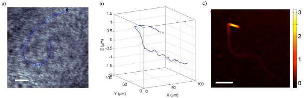
In addition to showing the sperm’s movement and behavior in real time, the novel method simultaneously provides detailed 3-D imaging of the sperm’s form and structure to detect potential infertility-causing anomalies, such as the “bent tail” that prevents the cells from swimming straight.
[ Also Read: Walnuts Improve Sperm Quality In Men: Study ]
The researchers say this is the first technique for collecting data on sperm cell motility—a key predictor of IVF success—in three dimensions and over time. They describe their method in a paper published Tuesday in The Optical Society’s (OSA) open-access journal Biomedical Optics Express.
Currently, sperm concentration and mobility in semen are assessed either by subjective visual evaluation or a process known as computer-assisted sperm analysis (CASA).
While the latter provides more detail and fewer errors than the former, CASA still only allows tracking and imaging in two dimensions.
In their new technique, the team of researchers from Italy and Belgium combined microscopy and holography—the creation of 3-D images—to visualize live sperm in not only two dimensions (the x and y positions) but according to their depth (z position) as well.
[ Also Read: Sky Selects Ultra-D Glasses-Free 3D Screens ]
And, “by acquiring a video of the moving sperm in 3-D, we add a fourth dimension – time,” said lead author Giuseppe Di Caprio of the Institute for Microelectronics and Microsystems of the National Research Council (NRC) in Naples, Italy, and Harvard University in Cambridge, Mass.
To achieve their new tracking system, the researchers first separated laser light into two beams. They transmitted one beam through a dish containing live, swimming sperm cells and then recombined it, after magnification through a microscope, with the second beam.
Di Caprio says that the 3-D imaging technique, known formally as digital holographic microscopy (DHM), yields morphology and motility data on sperm consistent with that found in previous studies, but with the unprecedented bonus of seeing cause and effect relationships between the two.
Founded in 1916, The Optical Society (OSA) is the leading professional society for scientists, engineers, students and business leaders who fuel discoveries, shape real-world applications and accelerate achievements in the science of light.
In the picture above: The track of a single sperm cell with a bent tail, in two dimensions (left) and three dimensions (right). Credit: Biomedical Optics Express.
Support RMN News Service for Independent Fearless Journalism. Scan the following QR Code to Donate.

In today’s media world controlled by corporates and politicians, it is extremely difficult for independent editorial voices to survive. Raman Media Network (RMN) News Service has been maintaining editorial freedom and offering objective content for the past 15 years despite enormous pressures and extreme threats. In order to serve you fearlessly in this cut-throat world, RMN News Service urges you to support us financially with your donations. You may please click here and choose the amount that you want to donate. Thank You. Rakesh Raman, Editor, RMN News Service.