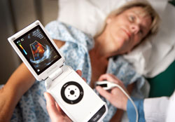
Scripps Study on Pocket Ultrasound Device
Nearly 200 years after the introduction of the stethoscope, the accuracy of a pocket ultrasound device that enables a physician to “look” at a patient’s heart during routine physical exams has been validated for the first time in peer-reviewed research led by Scripps Translational Science Institute (STSI) and Scripps Health.
Roughly the size of a smart phone, the Vscan pocket ultrasound used for point of care assessment of heart health could significantly reduce costs from traditional echocardiograms and improve the quality of care. Research was published in the July 5 issue of the Annals of Internal Medicine.
[ Also Read: IBM Study on Connected Health Devices ]“Pocket echos used during physical examinations may have the potential to reduce the number of unnecessary echocardiograms, particularly when used by a clinician trained in obtaining and interpreting the images,” said Dr. Eric J. Topol, cardiologist at Scripps Clinic, chief academic officer at Scripps Health, director of Scripps Translational Science Institute and principal investigator on the study.
“Approximately 20 million echocardiograms are conducted in the U.S. every year, each costing $1,500 or more and requiring a return appointment for a hospital or clinic echo laboratory for an extended session of about 45 minutes. A pocket echocardiogram could significantly reduce costs and improve the quality of the patient experience.”
Research showed that Vscan provided accurate assessments of ejection fraction, a measurement of how well the heart is pumping, and other measures for assessing heart health in patients.
[ Also Read: Medicines for Diseases that Strike Women ]In the study, physicians evaluated the hearts of 97 patients using the Vscan pocket ultrasound device to visually assess various heart structures within a five-minute timeframe in order to simulate the length of time of a physical examination.
Conclusions drawn from these imaging results were compared to conclusions drawn from imaging results also obtained from standard transthoracic echocardiography (TTE) machines.
The study evaluated the ability of the observer to visualize heart structures as well as accuracy in interpreting the images. The study also calculated difference in accuracy between cardiology attending physicians and less experienced cardiology fellows with two months or less training in echocardiographic interpretation.
[ Also Read: Go Red for Women with Love for Their Heart ]In the study, pocket ultrasound images were adequate for visualizing ejection fraction 95 percent of the time, wall motion abnormality 83 percent of the time, left ventricular end-diastolic dimension 95 percent of the time, pericardial effusion 94 percent of the time, mitral valve 90 percent of the time, aortic valve 82 percent of the time, and inferior vena cava (the large vein carrying blood to the heart) size 75 percent of the time.
The study says accuracy of interpretation of pocket ultrasound images was highest when assessing ejection fraction and aortic valve, and lowest when assessing inferior vena cava size. Accuracy and between-physician agreement was higher for experienced cardiologists than for less experienced cardiology fellows.


